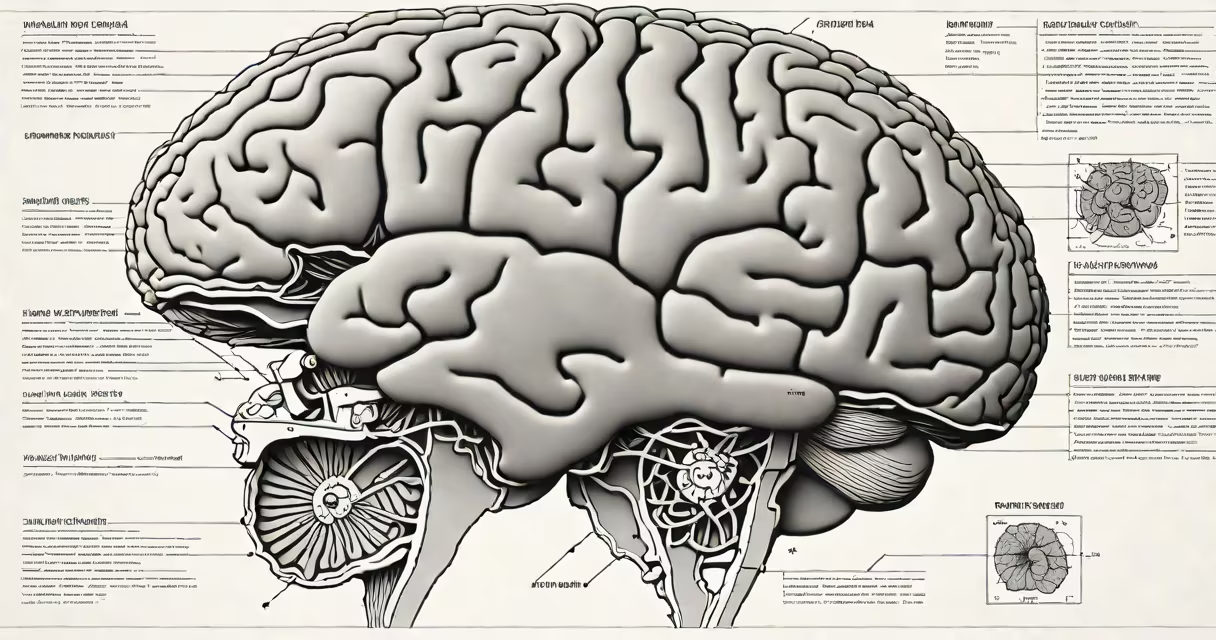
Human brain MRI anatomy uses magnetic resonance imaging to visualize the brain's internal structure, including gray matter, white matter, cerebrospinal fluid-filled ventricles, and critical regions such as the hippocampus and amygdala. The technique relies on magnetic fields and radio waves to generate high-resolution images without ionizing radiation.
MRI scans produce detailed cross-sectional views of the brain by detecting differences in water content and tissue density. These variations create contrast between gray matter (neuron cell bodies), white matter (myelinated axons), and cerebrospinal fluid. A typical scan captures hundreds of slices through the brain, enabling clinicians and researchers to map anatomical features with sub-millimeter precision.
For clinical diagnosis, MRI identifies abnormalities such as tumors, strokes, multiple sclerosis lesions, and developmental anomalies. In research, it supports studies on brain function, aging, and neurological disorders. The technique is safe, widely available, and forms the foundation of modern neuroimaging.
MRI operates by aligning hydrogen protons in water molecules within the brain using a useful magnetic field. Radiofrequency pulses then disturb this alignment. When the protons return to equilibrium, they emit signals detected by coils around the head. A computer reconstructs these signals into detailed images.
Different pulse sequences show specific tissue properties. T1-weighted scans emphasize anatomy-gray matter appears darker than white matter. T2-weighted scans show fluid-rich areas, making ventricles and edema visible. FLAIR (Fluid-Attenuated Inversion Recovery) suppresses cerebrospinal fluid signal, improving lesion detection near ventricles.
Functional MRI (fMRI) measures blood flow changes linked to neural activity. While not structural, fMRI often overlays onto structural MRI scans to pinpoint active brain regions during tasks like memory recall or decision-making. This integration allows researchers to correlate brain function with anatomy.
The following table outlines major anatomical features visible in standard MRI scans:
| Structure | Appearance on MRI | Function |
|---|---|---|
| Cerebral cortex | Thin outer layer; gray matter, folded into gyri and sulci | Higher cognition, sensory processing, motor control |
| Basal ganglia | Deep clusters of gray matter; appear dark on T1 | Movement coordination, habit formation |
| Hippocampus | Curved structure in temporal lobe; slightly darker than surrounding tissue | Memory consolidation, spatial navigation |
| Thalamus | Central relay station; symmetric oval structures | Sensory and motor signal relay to cortex |
| Ventricles | Dark, fluid-filled spaces; especially prominent on T2/FLAIR | Contain cerebrospinal fluid; cushion brain |
These structures are consistently identifiable across individuals using standardized protocols. For example, the hippocampus typically measures 3.5-4.5 cm in length and 1.5-2.0 cm in width in adults. Its volume decreases by about 0.5% per year after age 60, a trend observed in longitudinal MRI studies.
Many assume MRI shows brain activity directly. It does not. MRI detects structural differences or blood flow changes related to neural activity. fMRI measures blood oxygen level-dependent (BOLD) signals, which reflect metabolic demand-not electrical firing.
Another confusion arises between MRI and CT scans. CT uses X-rays and is faster, but exposes patients to ionizing radiation. MRI avoids radiation and provides superior soft-tissue contrast. However, CT remains preferred in trauma cases due to speed and availability in emergency settings.
A lesser-known fact: MRI scans are sensitive to motion. Even small head movements during acquisition cause blurring or ghosting artifacts. To minimize this, patients lie still for 20-45 minutes while the scanner runs sequences. Some centers use real-time motion correction algorithms to improve image quality.
In a 2021 study published in Nature Neuroscience, researchers used high-field 7T MRI to map cortical layers in living humans. They identified six distinct layers in the primary visual cortex, confirming findings from postmortem histology. This demonstrated that MRI can resolve microstructural details previously thought unattainable without invasive methods.
Consider a patient presenting with memory loss. A neurologist orders a brain MRI. The scan reveals bilateral hippocampal atrophy-volume reduced to 30% of normal size. This finding supports a diagnosis of early-stage Alzheimer's disease, confirmed later by cerebrospinal fluid biomarkers and cognitive testing.
Another example: a 32-year-old woman with epilepsy undergoes a 3T MRI. The scan detects a small cortical malformation in the left temporal lobe-abnormal gray matter folding and increased T2 signal. This lesion correlates with seizure onset zones mapped via EEG. Surgical removal improves outcomes.
People frequently mistake MRI images for actual brain activity maps. A common error is interpreting colorful activation patterns from fMRI as direct evidence of thoughts or emotions. In reality, those colors represent statistical significance of blood flow changes, not mental states.
Also, MRI does not distinguish between healthy and diseased tissue in all cases. For example, a glioma may mimic normal white matter on T1-weighted scans. Contrast agents like gadolinium are often required to improve tumor visibility. Without them, small lesions can be missed.
Even experienced clinicians sometimes misidentify normal anatomical variants. For instance, the arachnoid cyst-a benign fluid-filled space between brain membranes-can resemble a tumor on initial view. Confirmatory imaging or follow-up scans are needed to avoid unnecessary interventions.
I once reviewed a series of MRI scans from a university clinic. One patient had a large, asymmetrical lateral ventricle on the right side. Initial interpretation suggested hydrocephalus. But upon reviewing axial and coronal planes, we noticed the asymmetry was due to normal variation in cerebral spinal fluid distribution. No treatment was needed.
Human brain MRI anatomy provides precise visualization of brain structure using magnetic resonance imaging. It enables accurate diagnosis of neurological conditions, guides surgical planning, and supports neuroscience research. Understanding its capabilities and limitations prevents misinterpretation and ensures proper use in clinical and scientific contexts.