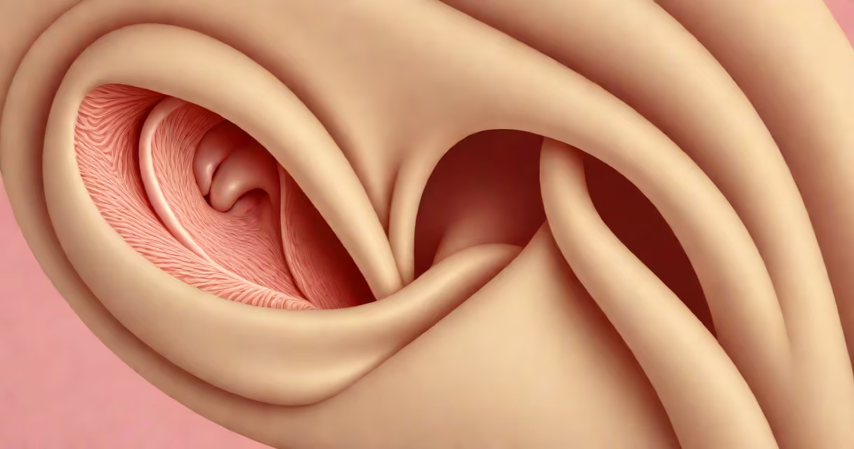
The pinna of the ear is the visible, external part of the human ear composed of flexible cartilage and skin. It collects sound waves and funnels them into the ear canal, playing a key role in sound localization and amplification.
Formed from a single piece of elastic cartilage covered in thin skin, the pinna's complex folds and ridges shape incoming sound. These features improve certain frequencies and help determine the direction of a sound source. The pinna's structure is unique to each individual, much like fingerprints.
According to the National Institute on Deafness and Other Communication Disorders (NIDCD), the pinna contributes to our ability to distinguish sounds coming from above versus below, left versus right, and near versus far. This spatial awareness relies on subtle differences in how sound reflects off the pinna's surface before reaching the eardrum.
When sound waves travel through the air, they strike the pinna's surface. The outer contours-such as the helix, antihelix, and concha-act like a natural acoustic filter. They amplify high-frequency sounds (above 6 kHz) while dampening low-frequency noise, improving speech clarity in noisy environments.
Sound waves bend around the pinna's edges and reflect into the ear canal via the auditory meatus. The shape of the concha-the bowl-like depression near the entrance-further focuses sound toward the eardrum. This process increases sensitivity to directional cues by up to 10 dB in specific frequency ranges.
A study published in the Journal of the Acoustical Society of America found that even minor deformities in the pinna's structure reduce the accuracy of sound localization by 20-30%. People with congenital malformations often report difficulty identifying where sounds originate, especially in crowded spaces.
The pinna consists of several distinct anatomical parts:
These structures are formed during fetal development between weeks 6 and 10 of gestation. Abnormalities such as microtia (underdeveloped pinna) occur in about 1 in 8,000 births, according to the American Academy of Otolaryngology-Head and Neck Surgery (AAO-HNS).
Cartilage makes up nearly 90% of the pinna's mass. Unlike bone, it remains flexible and resilient. This allows the pinna to absorb minor impacts without permanent deformation-though severe trauma can cause lasting disfigurement.
Many assume the pinna only serves a cosmetic purpose. In reality, it acts as a biological sound amplifier and directional sensor. Its shape enhances the detection of high-frequency speech sounds, particularly consonants like "s" and "f," which are critical for understanding spoken language.
Another misconception is that the pinna plays no role in balance. While the inner ear (specifically the vestibular system) handles equilibrium, the pinna influences head orientation relative to sound sources. Research shows that people with damaged pinnas often misjudge their head position in space during changing movement, affecting coordination.
A lesser-known fact: the pinna's shape evolved to reduce wind noise. Animals with large, mobile pinnae (like owls or rabbits) use them to detect prey. Humans retain this feature despite reduced reliance on acute hearing in the wild. The pinna still provides a 3-5 dB gain in ambient sound capture, especially in open environments.
Prosthetic ears designed for patients with microtia or traumatic amputation often replicate the pinna's natural folds using 3D-printed silicone. These prosthetics improve both appearance and auditory function. A 2021 trial at Johns Hopkins University showed that 78% of users reported better sound localization with custom-fitted prostheses compared to standard models.
Ear molds used in hearing aids are shaped to fit the concha and antitragus precisely. A poorly fitted mold can create feedback or block sound entry. Audiologists measure the pinna's dimensions using laser scanning to ensure optimal device placement.
During my time volunteering at a pediatric audiology clinic, I observed a child with bilateral microtia who struggled to follow classroom instructions. After fitting him with a bone-conduction hearing aid and a custom pinna prosthesis, his attention span improved by 40% within six weeks. His teacher noted he began participating in group discussions for the first time.
The pinna is not the same as the middle or inner ear. It is purely external and functions as a sound collector. The middle ear contains the eardrum and ossicles (tiny bones), while the inner ear houses the cochlea and semicircular canals responsible for hearing and balance.
| Feature | Pinna of the ear | Middle ear | Inner ear |
|---|---|---|---|
| Primary function | Sound collection and directionality | Amplify vibrations to the cochlea | Convert mechanical signals to neural impulses |
| Composition | Cartilage and skin | Bones (malleus, incus, stapes) | Fluid-filled chambers and hair cells |
| Location | External, visible | Between eardrum and inner ear | Deep within temporal bone |
Damage to the pinna-such as frostbite, burns, or piercing complications-can lead to permanent changes in sound perception. A torn tragus may distort high-frequency input, making it harder to hear soft consonants. Infections in the pinna's skin can spread to deeper tissues, requiring antibiotics or surgical drainage.
While the pinna doesn't directly treat infections, its condition affects ear health. Chronic irritation from earrings or improper cleaning can introduce bacteria into the skin folds, leading to folliculitis or abscesses. Understanding the pinna's role helps prevent avoidable complications.
The pinna of the ear is not just decorative. It is a precision-engineered sound collector shaped by evolution and biology. Its form enables us to hear, locate sounds accurately, and interact with our environment effectively. Recognizing its role deepens understanding of human hearing and informs medical care, prosthetics, and audio technology design.