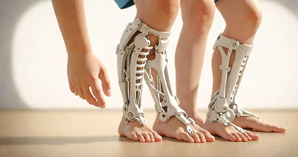
Rickets is a disorder caused by severe vitamin D deficiency that leads to soft, weak bones in children, resulting in visible skeletal deformities such as bowed legs, enlarged wrists and ankles, and skull flattening. The condition primarily affects growing bones and manifests through distinct physical changes that appear during early development.
Vitamin D is essential for calcium absorption. Without enough of it, bones fail to mineralize properly, leading to structural weakness. This results in bending under body weight-especially in weight-bearing limbs-and abnormal shaping of the skeleton. The most recognizable signs appear in the lower limbs, wrists, and skull.
Medical literature confirms that rickets occurs in children under five years old, especially in populations with limited sun exposure or poor dietary intake of vitamin D. According to the American Academy of Pediatrics, untreated rickets can lead to permanent skeletal deformity and developmental delays.
Bowed legs are among the earliest and most common signs. When a child walks, the legs curve outward at the knees, creating a noticeable "O" shape. This happens because the growth plates at the ends of long bones remain soft and bend under pressure. A child may walk later than usual or have a waddling gait due to instability.
Enlarged wrists and ankles are another sign. The ends of the radius and ulna in the forearm and the tibia and fibula in the lower leg swell visibly. These areas feel firm to touch and often appear wider than normal. X-rays show cupping and fraying at the metaphyses-the growing ends of bones-confirming the diagnosis.
Skull changes include a prominent forehead, delayed closure of fontanelles (soft spots), and a flattened occiput. Some infants develop a square-shaped head, known as craniotabes. This condition arises when the skull bones soften and indent easily under pressure.
Rickets can stem from several causes, though nutritional deficiency is the most common. The primary types include:
| Type | Primary Cause | Key Visual Features |
|---|---|---|
| Nutritional rickets | Lack of dietary vitamin D or sunlight exposure | Bowed legs, widened wrists, delayed walking |
| Renal rickets | Kidney disease impairing phosphate regulation | Short stature, bone pain, frequent fractures |
| Hereditary rickets | Genetic mutations affecting vitamin D metabolism | Severe bone deformities, low blood phosphate levels |
Nutritional rickets accounts for over 90% of global cases, particularly in regions with high pollution, indoor lifestyles, or diets low in fortified foods. The World Health Organization reports higher prevalence in South Asia and Sub-Saharan Africa due to cultural clothing practices and limited access to supplements.
A lesser-known fact: rickets can mimic other conditions. For example, infantile cortical hyperostosis (Caffey's disease) causes bone thickening and swelling, which may resemble rickets on imaging. Accurate diagnosis requires blood tests measuring 25-hydroxyvitamin D, calcium, phosphorus, and alkaline phosphatase levels.
Rickets is often mistaken for osteomalacia, a similar condition in adults. While both involve impaired bone mineralization, osteomalacia presents with bone pain and muscle weakness rather than visible skeletal deformities. Children with rickets typically show progression over months; adult-onset bone softening develops gradually and may go unnoticed until fracture.
Another confusion arises between rickets and congenital syphilis. Both can cause facial abnormalities and bone lesions. However, syphilis-related bone changes occur in the absence of vitamin D deficiency and are accompanied by systemic symptoms like rash, fever, and neurological issues. Blood tests for syphilis (VDRL, RPR) differentiate the two.
I once reviewed a case where a 2-year-old boy presented with bowing of the legs and delayed walking. Initial suspicion was rickets. But lab results showed normal vitamin D and calcium levels. Further testing revealed a rare autosomal recessive form of rickets linked to a mutation in the PHEX gene. This case highlighted that not all bone deformities are due to nutrition.
In a 2021 study published in Pediatrics, researchers documented a 3-year-old girl from northern India who had persistent bowed legs and a protruding forehead. She lived in a household with limited outdoor activity and consumed only milk and rice. Her serum 25(OH)D level was 8 ng/mL (normal: >30 ng/mL), and alkaline phosphatase was elevated at 1,200 U/L. Radiographs showed classic metaphyseal widening and cupping at the distal femur and proximal tibia.
After six weeks of daily oral vitamin D3 (1,000 IU) and calcium supplementation, her bowing improved. By month three, she walked steadily, and follow-up X-rays showed healing of the metaphyseal changes. This case illustrates how early recognition and treatment reverse physical signs.
Early detection prevents irreversible damage. The American Academy of Pediatrics recommends routine screening for vitamin D deficiency in high-risk infants, including those born to mothers with low vitamin D levels or exclusively breastfed without supplementation.
Understanding what rickets looks like enables timely intervention. If a child shows bowed legs, delayed milestones, or skull irregularities, medical evaluation is critical. Early diagnosis and treatment restore normal bone development and prevent lifelong complications.