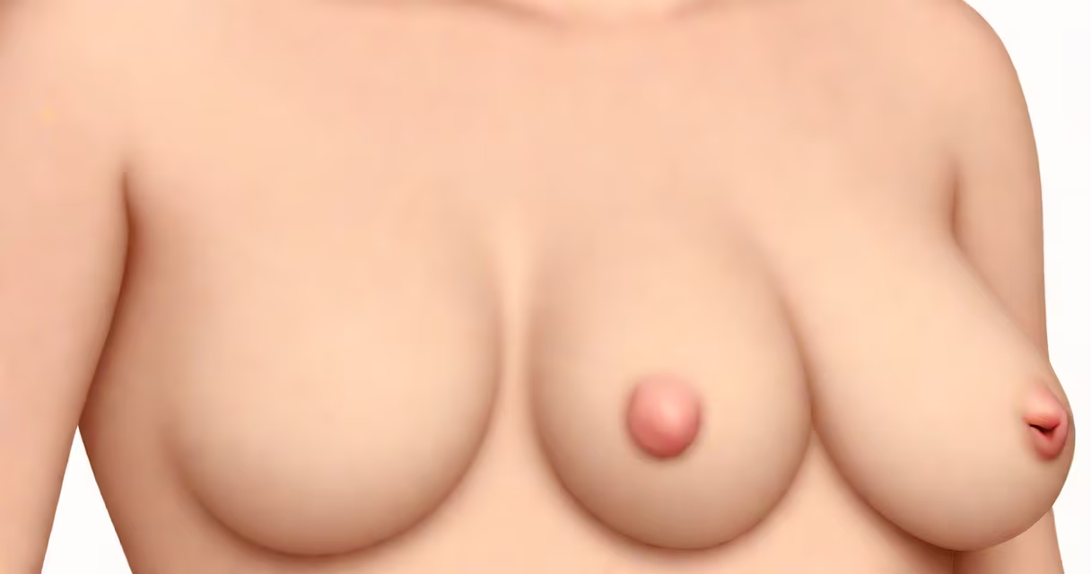
A lump behind the nipple in women typically results from a blocked milk duct, a cyst, or an infection such as mastitis. While most are benign, any new lump requires clinical evaluation to exclude breast cancer.
These growths form in the mammary ducts or glands behind the nipple. They often appear as firm, movable nodules under the skin. The area may feel tender, especially during hormonal shifts like menstruation or pregnancy. Not all lumps cause pain, but changes in size, texture, or associated nipple discharge demand attention.
According to the American Cancer Society, about 1 in 8 U.S. women will develop invasive breast cancer over their lifetime. Early detection through physical exams and imaging reduces mortality. A lump behind the nipple is not a guaranteed sign of cancer, but it must be assessed promptly.
Most lumps behind the nipple originate in the ductal system. Milk ducts extend from the base of the nipple toward the chest wall. When a duct becomes obstructed-by thickened secretions, scar tissue, or inflammation-it can swell and form a palpable mass. These are often called intraductal papillomas when they grow inside the ducts.
Cysts are fluid-filled sacs that develop in breast tissue. They're common in women aged 35-50 and may fluctuate in size with the menstrual cycle. Ultrasound can distinguish a cyst from a solid mass. Simple cysts are almost always harmless, but complex ones require biopsy.
Chronic inflammation, such as from recurrent mastitis, can lead to fibrosis-scar-like tissue that feels firm. This tissue forms in response to injury or infection and may persist even after healing. In rare cases, a lump may represent a benign tumor like a fibroadenoma or a malignant tumor like ductal carcinoma in situ (DCIS).
Not all lumps behind the nipple are the same. The following table outlines key types based on origin, appearance, and likelihood of malignancy:
| Type | Origin | Appearance | Benign Risk | Diagnostic Tool |
|---|---|---|---|---|
| Blocked milk duct | Ductal obstruction | Firm, round, sometimes tender | Very high | Ultrasound, clinical exam |
| Cyst | Fluid accumulation | Soft, fluctuant, may shrink | Extremely high | Ultrasound, aspiration |
| Intraductal papilloma | Overgrowth in duct lining | Small, may cause bloody discharge | High, but can mimic cancer | Mammogram, MRI, biopsy |
| Fibroadenoma | Connective tissue overgrowth | Smooth, mobile, rubbery | Very high | Ultrasound, biopsy if large |
| DCIS (ductal carcinoma in situ) | Abnormal cells in ducts | Often asymptomatic, detected via screening | Low unless untreated | Mammogram, biopsy |
Many assume any lump behind the nipple is cancer. That's incorrect. The National Breast Cancer Foundation reports that over 80% of breast lumps are noncancerous. However, ignoring a new lump increases risk of delayed diagnosis. A lump that persists beyond one menstrual cycle or grows larger should be evaluated.
Another misconception is that only older women get breast issues. In reality, younger women-especially those with dense breast tissue-can develop lumps. A 2022 study in JAMA Oncology found that breast cancer incidence in women under 40 rose 1.6% annually between 2000 and 2019.
Any new lump behind the nipple warrants a visit to a healthcare provider. Red flags include:
I once examined a 34-year-old patient who reported a small lump behind her left nipple. She had noticed it three weeks prior and assumed it was hormonal. On exam, it was firm, non-tender, and didn't move with pressure. An ultrasound revealed a 1.2 cm solid mass. Biopsy confirmed a low-grade phyllodes tumor-a rare but benign fibroepithelial growth. Removal prevented future complications.
Imaging protocols depend on age and risk. Women under 40 with dense breasts often start with ultrasound. Those over 40 typically undergo mammography. If a lesion appears suspicious, MRI or biopsy follows. The American College of Radiology recommends screening mammograms every 1-2 years for average-risk women starting at age 40.
People often mistake a lump behind the nipple for a hernia or lymph node swelling. Hernias occur in the abdominal wall and are felt near the groin or lower abdomen. Lymph nodes in the armpit may swell due to infection but don't typically lie directly behind the nipple.
Another confusion arises with gynecomastia-breast enlargement in men. Though rare in women, some may confuse it with a lump. Gynecomastia involves diffuse breast tissue growth, not a localized mass. A lump behind the nipple is usually discrete and distinct from surrounding tissue.
A lump behind the nipple is not the same as a subareolar abscess. Subareolar abscesses form under the nipple and are caused by bacterial infection. They present with fever, severe pain, and pus drainage. Treatment includes antibiotics and drainage. A lump behind the nipple may be related but is not necessarily infected.
One non-obvious fact: nipple discharge can precede a lump. A 2021 study in Annals of Surgical Oncology found that 37% of women diagnosed with DCIS reported unilateral nipple discharge before imaging detected the lesion. Bloody or serous discharge should never be ignored.
If a lump is discovered, schedule a clinical breast exam within four weeks. Your doctor may recommend imaging-ultrasound for younger women, mammography for those over 40. If the lesion is suspicious, a biopsy is performed. Core needle biopsy is the gold standard, with a 95% accuracy rate in distinguishing benign from malignant tissue.
For benign findings, follow-up is typically every 6-12 months. For persistent or growing masses, surgical excision may be advised. In rare cases, a lump behind the nipple can be an early sign of Paget's disease of the breast-an aggressive form of ductal cancer affecting the nipple skin. Symptoms include itching, scaling, and ulceration.