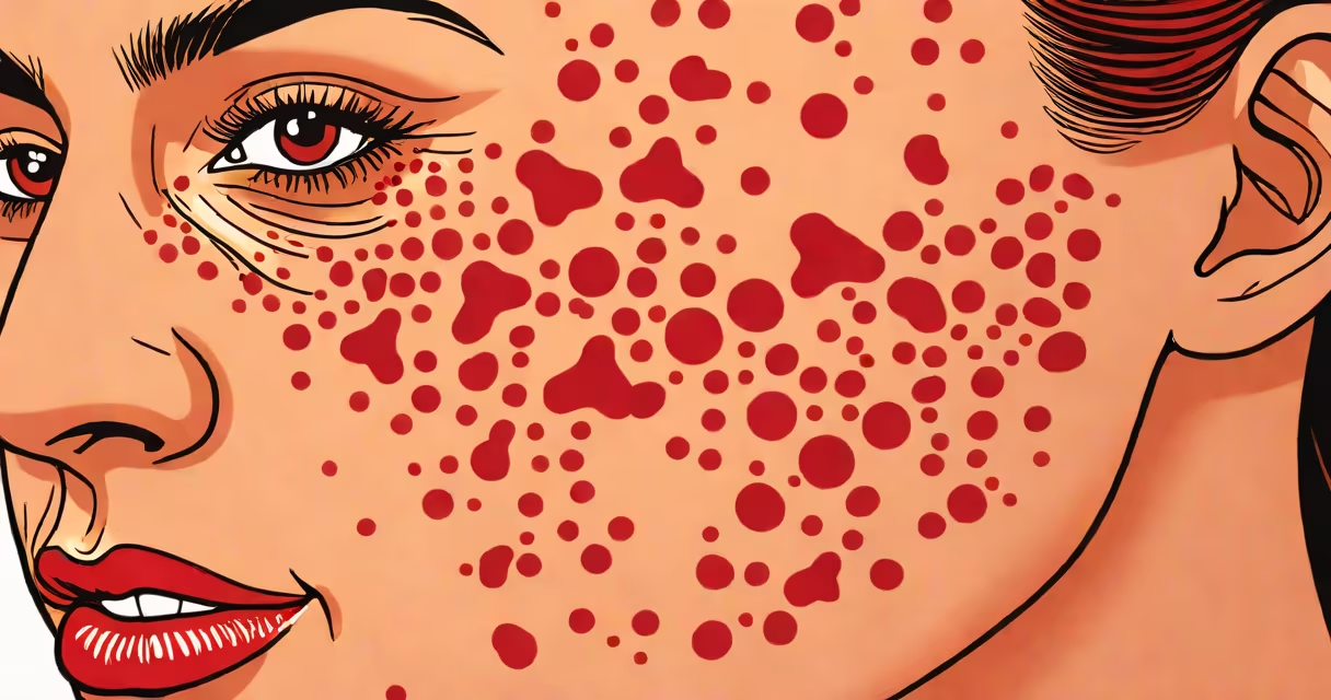
Discoid lupus erythematosus (DLE) results from an autoimmune reaction where the body attacks its own skin cells, primarily triggered by ultraviolet (UV) radiation and genetic predisposition. The condition manifests as persistent, scaly, red patches-often on the face, ears, and scalp-that can scar and cause permanent hair loss.
DLE is a chronic form of cutaneous lupus, distinct from systemic lupus erythematosus (SLE), though 10-20% of DLE patients develop SLE over time. Autoantibodies such as anti-Ro/SSA and antinuclear antibodies (ANA) are frequently detected in blood tests, supporting the autoimmune basis. The immune system mistakenly identifies healthy skin cells as foreign, leading to inflammation and tissue damage.
UV light exposure is the most consistent environmental trigger. A 2021 study published in JAMA Dermatology found that 78% of DLE flare-ups correlated with recent sun exposure or high UV index days. Sunlight activates dendritic cells in the skin, which then present self-antigens to T cells, initiating the inflammatory cascade. This explains why lesions typically appear on sun-exposed areas like the nose, cheeks, and forehead.
The process begins when UV radiation alters proteins in keratinocytes-the primary skin cells. These altered proteins are processed and displayed on the cell surface, attracting autoreactive CD4+ T cells. Once activated, these T cells release cytokines like interferon-alpha and tumor necrosis factor, amplifying inflammation and recruiting macrophages and neutrophils.
As immune cells infiltrate the dermis, they degrade collagen and disrupt the basement membrane. This leads to epidermal atrophy, hyperkeratosis (thickened outer layer), and fibrosis-scarring that replaces normal skin. The resulting lesions are well-demarcated, red, raised plaques with adherent scale. Over time, they may shrink and leave hypopigmented or atrophic scars.
A 2019 longitudinal analysis from the National Institutes of Health (NIH) tracked 187 DLE patients over five years. Of those, 13% developed SLE, defined by the presence of at least four of the 11 American College of Rheumatology (ACR) criteria, including renal involvement or positive anti-dsDNA antibodies. This underscores the importance of monitoring DLE patients for systemic signs.
While UV light is the strongest external trigger, several internal and environmental factors contribute to onset and flares:
Despite common belief, DLE is not contagious or infectious. It cannot be passed through contact. The misconception likely stems from the visible rash resembling fungal infections or psoriasis. However, histopathology shows interface dermatitis with lymphocytic infiltration-distinct from infectious patterns.
Many assume DLE is simply a severe form of eczema or psoriasis due to similar scaling. But unlike these conditions, DLE lesions show a characteristic "follicular plugging" under the microscope-clogged hair follicles filled with keratin. This feature is absent in psoriasis and eczema.
Another myth is that DLE always progresses to systemic disease. In reality, most cases remain localized. A 2022 review in Autoimmunity Reviews reported that 80-90% of DLE patients never develop SLE. The risk increases with ANA positivity, anti-Ro/SSA antibodies, and early-onset disease.
Some patients mistake DLE for rosacea or seborrheic dermatitis. Rosacea features flushing, telangiectasia, and papules, but lacks scarring and annular lesions. Seborrheic dermatitis affects oily zones like the scalp and nasolabial folds, but presents with greasy yellow scales-not thick, red plaques.
Diagnosis relies on clinical appearance, biopsy, and serology. A punch biopsy showing lymphocytic infiltration at the dermo-epidermal junction, colloid bodies (apoptotic keratinocytes), and fibrosis confirms DLE. Direct immunofluorescence may reveal IgM and C3 deposits along the basement membrane.
Consider the case of a 42-year-old woman presenting with a 6-month history of a red, scaly patch on her left cheek. She reported worsening after beach visits. Skin exam revealed a well-circumscribed, hyperpigmented plaque with central atrophy and peripheral scale. Biopsy confirmed interface dermatitis with follicular plugging. ANA was positive at 1:160, anti-Ro/SSA positive. She was diagnosed with DLE and prescribed hydroxychloroquine and strict sun protection.
After six months of daily broad-spectrum sunscreen (SPF 50+), avoidance of peak sun hours, and daily hydroxychloroquine 200 mg, the lesion regressed by 70%. This illustrates how targeting both UV exposure and immune modulation improves outcomes.
Understanding differences between lupus subtypes clarifies DLE's unique profile:
| Type | Primary Site | Scarring Risk | Systemic Involvement | Antibody Association |
|---|---|---|---|---|
| Discoid lupus (DLE) | Face, scalp, ears | High (up to 50%) | Low (10-20%) | Anti-Ro/SSA, ANA |
| Subacute cutaneous lupus (SCLE) | Trunk, arms, neck | Low | Moderate (joint pain, fatigue) | Anti-Ro/SSA (high sensitivity) |
| Systemic lupus (SLE) | Multiple organs | Variable | High (kidneys, heart, lungs) | anti-dsDNA, anti-Smith |
Unlike SCLE, which presents with non-scarring, annular or psoriasiform lesions, DLE causes irreversible scarring. Unlike SLE, DLE rarely involves internal organs-but the risk of progression necessitates long-term follow-up.
During a clinic visit, I observed a patient with DLE who had lost all hair on the right side of his scalp due to scarring alopecia. He had ignored sun exposure for years. After starting hydroxychloroquine and wearing a wide-brimmed hat, new hair grew in the unaffected area. This highlights the preventable nature of DLE progression with proper management.